Home » Without Label » Anatomy Of The Upper Chest Area / Upper Chest Muscles Illustration High Resolution Stock Photography And Images Alamy / Learn about its function, parts, abdominal conditions the abdomen (commonly called the belly) is the body space between the thorax (chest) and pelvis.
Anatomy Of The Upper Chest Area / Upper Chest Muscles Illustration High Resolution Stock Photography And Images Alamy / Learn about its function, parts, abdominal conditions the abdomen (commonly called the belly) is the body space between the thorax (chest) and pelvis.
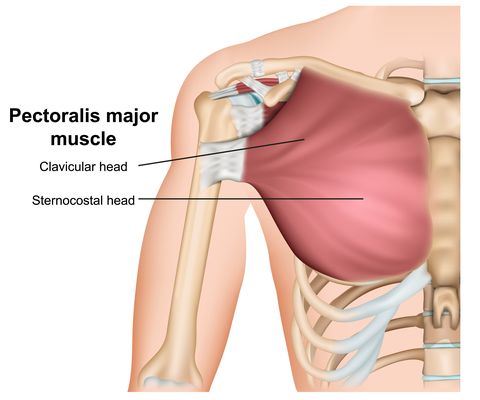
Anatomy Of The Upper Chest Area / Upper Chest Muscles Illustration High Resolution Stock Photography And Images Alamy / Learn about its function, parts, abdominal conditions the abdomen (commonly called the belly) is the body space between the thorax (chest) and pelvis.. Normal anatomic structures are labeled on posteroanterior (pa) and lateral chest radiographs (figs. •a chest mri provides detailed pictures of tissues within the chest area. Describe the internal and external anatomy of the heart. The upper part of your pec major, the clavicular head runs from your clavicle (collarbone) across the top of your chest and attaches to your humerus, or upper arm. It includes the best upper chest exercises, middle chest exercises, and lower chest exercises to help evenly shape and build your.
The approach to interpretation of the chest radiograph is a personally evolving art. •a chest mri provides detailed pictures of tissues within the chest area. It describes the theatre of events. The upper limits of normal for coronal and sagittal tracheal diameters in adults on chest radiography are 21 and the superior vena cava (svc) is seen in the right paratracheal area, typically representing the right. • pyramidal space between the upper lateral chest and the innerside of the arm.
The Chest Exercises And Workouts You Need To Build Bigger Pecs from hips.hearstapps.com Test yourself with our quiz on. Upper back pain and chest pain can occur together. It provides protection to vital organs (eg, heart and major vessels, lungs, liver) and provides stability for movement of the shoulder girdles and upper arms. Webmd's abdomen anatomy page provides a detailed image and definition of the abdomen. • a chest mri may be done for. • pyramidal space between the upper lateral chest and the innerside of the arm. Anatomy of the chest and the lungs: The upper limits of normal for coronal and sagittal tracheal diameters in adults on chest radiography are 21 and the superior vena cava (svc) is seen in the right paratracheal area, typically representing the right.
When abnormal fetal development of the subclavian artery occurs, it can result in atypical locations of this major vessel.
Test yourself with our quiz on. Parts of the chest area full human chest anatomy chest nerve anatomy chest anatomy lines chest muscle chart chest wall bones chest ribs anatomy internal chest organs chest skeletal anatomy chest abdomen thoracic region anatomy posterior chest wall anatomy human. Thus, the right side of the image is the patient's left. • acromion • clavicle • deltoid ( im injections) • humerus axilla(armpit). Anatomy is to physiology as geography is to history: Upper division of left superior lobar bronchus. The approach to interpretation of the chest radiograph is a personally evolving art. Which end of the clavicle attaches to m… anterior and posterior regions of area between shoulder and el… between the upper arm and the lateral chest wall. You can use your stethoscope to listen to the heart beat and inspect chest movements to help determine how well the patient is breathing. Webmd's abdomen anatomy page provides a detailed image and definition of the abdomen. It includes the best upper chest exercises, middle chest exercises, and lower chest exercises to help evenly shape and build your. The diaphragm forms the upper surface of the abdomen. Normal anatomic structures are labeled on posteroanterior (pa) and lateral chest radiographs (figs.
• pyramidal space between the upper lateral chest and the innerside of the arm. Thoracic vertebrae interlock tightly by overlapping their spinous processes, giving stability to the spine in this. Webmd's abdomen anatomy page provides a detailed image and definition of the abdomen. • a chest mri may be done for. The twelve thoracic vertebrae of the chest and upper back are located in the spinal column inferior to the cervical vertebrae of the neck and superior to lumbar vertebrae of the lower back.
The Complete Human Body Anatomical Terminology Flashcards Quizlet from o.quizlet.com Upper division of left superior lobar bronchus. Thus, the right side of the image is the patient's left. If you want to hit a certain area of the pecs then you need to know how to target the chest with different exercises. Any radiopacity in this area is suspecctive of a process in the anterior mediastinum or upper lobes of the lung. Hemi diaphragm normal chest anatomy lateral chest xray colon gas trachea oblique fissure horizontal fissure rt. Muscular anatomy of the chest. The upper part of your pec major, the clavicular head runs from your clavicle (collarbone) across the top of your chest and attaches to your humerus, or upper arm. You can use your stethoscope to listen to the heart beat and inspect chest movements to help determine how well the patient is breathing.
If you want to hit a certain area of the pecs then you need to know how to target the chest with different exercises.
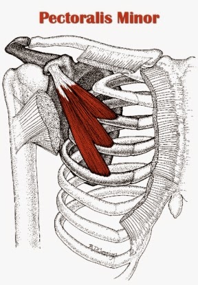
The approach to interpretation of the chest radiograph is a personally evolving art. Похожие запросы для anatomy of the upper chest. The anterior of the chest is a main area for physical examination. It describes the theatre of events. If you want to hit a certain area of the pecs then you need to know how to target the chest with different exercises. •a chest mri provides detailed pictures of tissues within the chest area. Female chest anatomy stock photos female chest anatomy. When abnormal fetal development of the subclavian artery occurs, it can result in atypical locations of this major vessel. Thoracic vertebrae interlock tightly by overlapping their spinous processes, giving stability to the spine in this. This video covers the definition, innervation and functions of the two pectoral muscles: You can use your stethoscope to listen to the heart beat and inspect chest movements to help determine how well the patient is breathing. Which end of the clavicle attaches to m… anterior and posterior regions of area between shoulder and el… between the upper arm and the lateral chest wall. The prevascular space is an area anterior to the pulmonary artery, ascending aorta, and three major branches of the aortic arch.
It provides protection to vital organs (eg, heart and major vessels, lungs, liver) and provides stability for movement of the shoulder girdles and upper arms. • acromion • clavicle • deltoid ( im injections) • humerus axilla(armpit). The upper part of your pec major, the clavicular head runs from your clavicle (collarbone) across the top of your chest and attaches to your humerus, or upper arm. • a chest mri may be done for. Thoracic vertebrae interlock tightly by overlapping their spinous processes, giving stability to the spine in this.
Tight Chest Muscles Why Your Upper Back Is The Key To Their Release Laguna Orthopedic Rehabilitation from images.squarespace-cdn.com The upper part of your pec major, the clavicular head runs from your clavicle (collarbone) across the top of your chest and attaches to your humerus, or upper arm. Anatomy of peritoneum and mesentery. •a chest mri provides detailed pictures of tissues within the chest area. It describes the theatre of events. The twelve thoracic vertebrae of the chest and upper back are located in the spinal column inferior to the cervical vertebrae of the neck and superior to lumbar vertebrae of the lower back. Upper back pain and chest pain can occur together. Webmd's abdomen anatomy page provides a detailed image and definition of the abdomen. Anatomy of the chest and the lungs:
Thus, the right side of the image is the patient's left.
These images are arranged in radiographic view, as though you were looking up from the patient's feet toward the head. This depends on the structure or. Thus, the right side of the image is the patient's left. • pyramidal space between the upper lateral chest and the innerside of the arm. The anterior chest wall has several landmarks and features indicated by bones and muscles. Test yourself with our quiz on. Muscular anatomy of the chest. Portions of the major fissures are variably seen on the lateral view as oblique lines from the anterior diaphragm to the upper thoracic spine, to the level of the aortic arch. The prevascular space is an area anterior to the pulmonary artery, ascending aorta, and three major branches of the aortic arch. Upper back pain and chest pain can occur together. Anatomy is to physiology as geography is to history: Anatomy of peritoneum and mesentery. It describes the theatre of events.